Leukoplakia is a white patch or plaque that develops on the mucous membranes of the oral cavity, commonly observed in smokers. It is considered a potentially precancerous lesion, often appearing on the tongue, inner cheeks, or floor of the mouth. Tobacco use, especially smoking, is a significant risk factor for leukoplakia due to the chronic irritation and exposure to carcinogens in tobacco smoke. Data from clinical studies indicate that smokers have a higher prevalence of leukoplakia compared to non-smokers. Histopathological analysis often reveals hyperkeratosis and epithelial dysplasia in these lesions, which may progress to oral cancer if untreated. Monitoring and early intervention are critical for smokers diagnosed with leukoplakia to prevent malignant transformation.
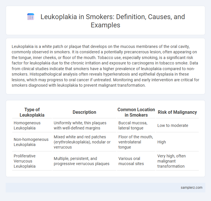
Table of Comparison
| Type of Leukoplakia | Description | Common Location in Smokers | Risk of Malignancy |
|---|---|---|---|
| Homogeneous Leukoplakia | Uniformly white, thin plaques with well-defined margins | Buccal mucosa, lateral tongue | Low to moderate |
| Non-homogeneous Leukoplakia | Mixed white and red patches (erythroleukoplakia), nodular or verrucous | Floor of the mouth, ventrolateral tongue | High |
| Proliferative Verrucous Leukoplakia | Multiple, persistent, and progressive verrucous plaques | Various oral mucosal sites | Very high, often malignant transformation |
Understanding Leukoplakia: Definition and Causes
Leukoplakia appears as white patches on the oral mucosa, commonly developing in smokers due to chronic irritation from tobacco smoke. This precancerous lesion results from abnormal keratinization triggered by harmful substances in cigarette smoke, increasing the risk of malignant transformation. Early identification and cessation of smoking are crucial to prevent progression to oral cancer.
The Link Between Smoking and Leukoplakia
Smoking is a primary risk factor for leukoplakia, a condition characterized by white patches on the mucous membranes of the mouth. Tobacco smoke contains carcinogens that irritate and damage the oral epithelium, promoting the development of precancerous lesions like leukoplakia. Studies show that smokers have a significantly higher incidence of leukoplakia compared to non-smokers, highlighting the strong link between smoking and the condition's onset.
Common Sites of Leukoplakia in Smokers
Leukoplakia in smokers commonly appears on the lateral borders of the tongue, the floor of the mouth, and the buccal mucosa. These sites are particularly vulnerable due to direct exposure to tobacco smoke and its carcinogenic components. Regular examination of these areas is essential for early detection and intervention.
Visual Characteristics of Leukoplakia Lesions
Leukoplakia lesions in smokers typically appear as thickened, white patches with irregular, well-defined borders on the oral mucosa, especially on the tongue, floor of the mouth, and inner cheeks. These plaques may exhibit a rough, fissured surface texture and are often asymptomatic, making early detection challenging. The lesions' persistent white coloration is due to keratin buildup, distinct from other mucosal changes caused by smoking.
Early Signs of Leukoplakia in Smokers
White or grayish patches appearing on the inner cheeks, gums, or tongue often signify early signs of leukoplakia in smokers. These lesions may feel slightly raised and cannot be easily scraped off, indicating abnormal cell growth linked to tobacco use. Persistent irritation from smoking increases the risk of these precancerous patches developing into oral cancer if left untreated.
Clinical Case Examples: Leukoplakia in Tobacco Users
A 52-year-old male smoker presented with a persistent white patch on the lateral border of the tongue, diagnosed clinically and histopathologically as leukoplakia. Tobacco use, particularly smoking, is a significant risk factor for this potentially premalignant lesion, which often appears as thickened, adherent, and non-removable plaques in the oral mucosa. Early detection and cessation of tobacco use are critical to prevent malignant transformation in tobacco-associated leukoplakia cases.
Risk Factors for Leukoplakia Progression
Chronic tobacco smoking significantly increases the risk of leukoplakia developing into oral cancer, with the risk amplifying in conjunction with alcohol consumption and poor oral hygiene. Persistent exposure to carcinogens in tobacco smoke leads to cellular mutations, making smokers more susceptible to malignant transformation of leukoplakic lesions. Early identification and cessation of smoking are critical to reducing the likelihood of leukoplakia progression in high-risk individuals.
Diagnostic Methods for Identifying Leukoplakia
Leukoplakia in smokers is primarily diagnosed through clinical examination and confirmed by biopsy, which helps distinguish it from malignant lesions. Toluidine blue staining aids in highlighting abnormal epithelial changes, while brush cytology serves as a non-invasive preliminary diagnostic tool. Advanced techniques like autofluorescence and narrow band imaging enhance early detection by identifying dysplastic tissue changes in the oral mucosa.
Treatment Options for Leukoplakia in Smokers
Treatment options for leukoplakia in smokers primarily include smoking cessation to reduce risk factors and promote lesion regression. Clinical management often involves regular monitoring and biopsy of suspicious areas to rule out malignant transformation. In certain cases, surgical excision, laser therapy, or topical agents like retinoids may be recommended to treat persistent leukoplakic patches.
Prevention and Lifestyle Changes for Smokers at Risk
Leukoplakia in smokers often appears as white patches on the tongue or inside the mouth, signaling potential precancerous changes. Preventive measures include quitting smoking, reducing alcohol consumption, and maintaining excellent oral hygiene to decrease the risk of malignant transformation. Regular dental check-ups and early detection play critical roles in managing leukoplakia and preventing progression to oral cancer.
example of leukoplakia in smoker Infographic