Xanthelasma is a medical condition characterized by yellowish plaques that typically appear on the eyelids, especially near the inner canthus. These deposits consist of cholesterol and lipid-laden macrophages and are often linked to underlying lipid metabolism disorders. Xanthelasma can serve as an early indicator of hyperlipidemia or other forms of dyslipidemia. Clinically, xanthelasma presents as soft, flat, or slightly elevated lesions that range from a few millimeters to several centimeters in size. The eyelid is the most common location, with the upper lids being more frequently affected than the lower lids. Diagnosis is usually clinical, supported by lipid profile testing to identify possible systemic lipid abnormalities.
Table of Comparison
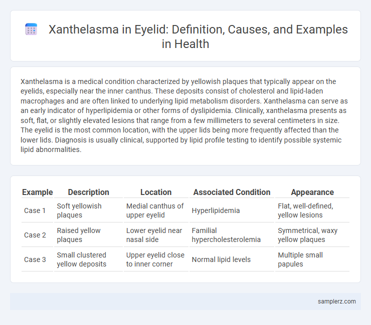
| Example | Description | Location | Associated Condition | Appearance |
|---|---|---|---|---|
| Case 1 | Soft yellowish plaques | Medial canthus of upper eyelid | Hyperlipidemia | Flat, well-defined, yellow lesions |
| Case 2 | Raised yellow plaques | Lower eyelid near nasal side | Familial hypercholesterolemia | Symmetrical, waxy yellow plaques |
| Case 3 | Small clustered yellow deposits | Upper eyelid close to inner corner | Normal lipid levels | Multiple small papules |
Understanding Xanthelasma: Definition and Causes
Xanthelasma are yellowish plaques that commonly appear on the eyelids due to lipid accumulation beneath the skin. These lesions are often linked to hyperlipidemia, where elevated cholesterol or triglyceride levels trigger fat deposits. Understanding the connection between lipid metabolism disorders and xanthelasma helps guide effective diagnosis and management.
Identifying Xanthelasma on the Eyelid
Xanthelasma on the eyelid appears as yellowish, flat, or slightly raised plaques typically located on the inner canthus or upper eyelid. These lipid-rich deposits often indicate underlying hyperlipidemia, necessitating lipid profile testing for accurate diagnosis. Early identification of xanthelasma helps prompt management to reduce cardiovascular risk associated with cholesterol abnormalities.
Common Characteristics of Eyelid Xanthelasma
Xanthelasma on the eyelids commonly presents as yellowish, soft, flat plaques typically located on the medial canthus of the upper eyelids. These lesions are often bilateral and symmetrical, reflecting lipid accumulation beneath the skin, primarily composed of cholesterol-laden foam cells. Associated factors include hyperlipidemia, diabetes mellitus, and advanced age, which contribute to the prevalence and recurrence of eyelid xanthelasma.
Clinical Example: Typical Presentation of Xanthelasma
Xanthelasma typically presents as soft, yellowish plaques located on the medial aspect of the eyelids, often bilateral and symmetrical. These lesions are usually painless, flat, and may gradually increase in size over time, commonly seen in middle-aged individuals. Clinical examination reveals lipid-laden macrophages within the dermis, which correlate with underlying dyslipidemia in many cases.
Xanthelasma vs. Other Eyelid Lesions
Xanthelasma presents as yellowish, soft, flat plaques on the eyelids, often near the inner canthus, distinct from other eyelid lesions such as chalazion, which is typically a firm, painless nodule, or basal cell carcinoma characterized by pearly, ulcerated nodules with telangiectasia. Unlike sebaceous cysts or papillomas, xanthelasma lesions are lipid-rich deposits linked to hyperlipidemia and are generally symmetrical and bilateral. Accurate diagnosis relies on clinical examination and lipid profiling to differentiate xanthelasma from malignant or infectious eyelid conditions.
Risk Factors Associated with Eyelid Xanthelasma
Xanthelasma on the eyelid is commonly linked to hyperlipidemia, with elevated low-density lipoprotein (LDL) cholesterol and triglyceride levels serving as primary risk factors. Other contributors include diabetes mellitus, hypertension, and advanced age, which increase the likelihood of lipid deposition in the periorbital skin. Genetic predisposition and lifestyle factors such as smoking and obesity further elevate the risk of developing eyelid xanthelasma.
Diagnosing Xanthelasma: What to Look For
Xanthelasma appears as yellowish, soft, flat plaques on the eyelids, commonly near the inner corners, indicating lipid deposits beneath the skin. Diagnosing xanthelasma involves a clinical examination focusing on the characteristic color, location, and texture of the lesions, often confirmed with lipid profile testing to identify underlying hyperlipidemia. Early detection is crucial to address potential cardiovascular risks associated with elevated cholesterol levels.
Case Study: Xanthelasma in an Adult Patient
Xanthelasma presents as yellowish, cholesterol-rich deposits on the eyelids, often signaling underlying lipid metabolism disorders. In a documented case study of an adult patient, the lesions appeared bilaterally on the upper eyelids, correlating with elevated LDL cholesterol and triglyceride levels. Targeted lipid-lowering therapy combined with surgical excision successfully reduced lesion size and prevented recurrence.
Treatment Options for Eyelid Xanthelasma
Treatment options for eyelid xanthelasma include surgical excision, laser therapy, chemical peels using trichloroacetic acid, and cryotherapy. Each method targets the cholesterol deposits differently, with surgical excision providing immediate removal and laser therapy reducing recurrence risk. Consultation with a dermatologist or ophthalmologist ensures selection of the most effective and safe treatment based on lesion size and patient skin type.
Preventing Xanthelasma Recurrence on Eyelids
Preventing xanthelasma recurrence on eyelids involves maintaining optimal cholesterol levels through a heart-healthy diet rich in omega-3 fatty acids and fiber. Regular monitoring of lipid profiles and managing underlying conditions such as hyperlipidemia and diabetes can reduce the risk of lipid deposits forming on the eyelids. Incorporating lifestyle changes, including consistent exercise and smoking cessation, supports long-term prevention of xanthelasma buildup.
example of xanthelasma in eyelid Infographic