A macule in measles appears as a flat, discolored spot on the skin, typically red or reddish-brown in color. These lesions usually measure less than 1 centimeter in diameter and are one of the earliest visible signs of measles infection. Macules often start on the face and neck, spreading downward to the rest of the body as the disease progresses. The presence of macules in measles is accompanied by other symptoms such as fever, cough, runny nose, and conjunctivitis. These flat spots do not raise above the skin surface, distinguishing them from papules or pustules found in other skin conditions. Monitoring the distribution and progression of macules helps healthcare providers confirm a measles diagnosis and evaluate the stage of the illness.
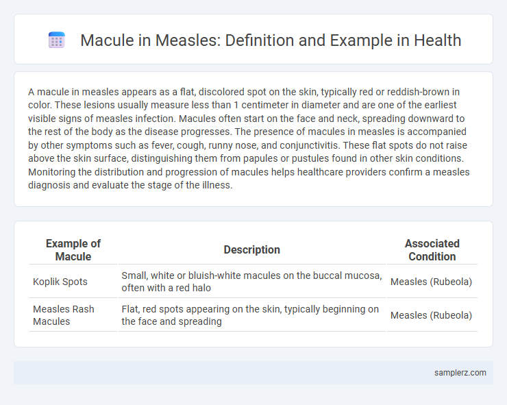
Table of Comparison
| Example of Macule | Description | Associated Condition |
|---|---|---|
| Koplik Spots | Small, white or bluish-white macules on the buccal mucosa, often with a red halo | Measles (Rubeola) |
| Measles Rash Macules | Flat, red spots appearing on the skin, typically beginning on the face and spreading | Measles (Rubeola) |
Definition of Macule in Measles
A macule in measles is a flat, discolored spot on the skin, typically red or pink, measuring less than 1 centimeter in diameter. It appears early in the disease as part of the characteristic rash, usually starting on the face and spreading downward. This non-raised lesion results from inflammation and blood vessel dilation caused by the measles virus infection.
Clinical Appearance of Measles Macules
Measles macules appear as flat, discolored spots on the skin, typically measuring 2-10 mm in diameter, with a characteristic reddish-brown hue that may coalesce into larger patches. These macules first emerge on the face and behind the ears before spreading downward to the trunk and limbs. The clinical appearance often includes Koplik spots inside the mouth, which are pathognomonic and help differentiate measles from other viral exanthems.
Common Locations of Measles Macules
Measles macules commonly appear first on the face, particularly around the hairline, forehead, and behind the ears, before spreading downward to the neck, trunk, and extremities. These flat, red spots typically measure 2 to 10 millimeters in diameter and may coalesce into larger irregular patches. The distribution pattern of measles macules helps distinguish this viral rash from other exanthems, aiding early diagnosis and effective management.
Progression and Evolution of Measles Macules
Measles macules begin as small, flat, red spots that typically appear 3 to 5 days after the onset of initial symptoms such as fever and cough. These macules rapidly coalesce and spread from the face downward to the trunk and limbs, evolving into larger, more confluent lesions within 24 to 48 hours. Over the course of 5 to 7 days, the rash fades in the order it appeared, often leaving behind temporary skin discoloration without scarring.
Differentiating Measles Macules from Other Lesions
Measles macules typically present as flat, red spots that appear 2-3 days after the onset of fever, often starting on the face and spreading downward. These lesions are distinct from papules or vesicles due to their flat nature and can be differentiated from rash caused by rubella or roseola by their larger size and confluence over time. The presence of Koplik spots inside the mouth serves as a key diagnostic marker to distinguish measles macules from other viral exanthems.
Pathophysiology Behind Measles Macules
Measles macules are the hallmark skin manifestation resulting from the virus-induced damage to epidermal cells and subsequent immune response. The pathophysiology involves the measles virus infecting keratinocytes and endothelial cells, triggering a local cytotoxic T-cell mediated reaction that leads to characteristic erythematous macular eruptions. Immune complex deposition and capillary dilation contribute to the development of these discrete, flat lesions typical in the prodromal phase of measles infection.
Diagnostic Importance of Macules in Measles
Macules in measles present as flat, red spots that are essential for early diagnosis due to their characteristic appearance and distribution on the face and trunk. Identifying these macules allows healthcare providers to differentiate measles from other viral exanthems and initiate timely isolation measures to prevent outbreaks. Early recognition of measles macules is critical for confirming infection through clinical assessment and supporting laboratory testing.
Associated Symptoms with Measles Macules
Measles macules appear as flat, red spots that often merge to form larger blotches on the skin. Associated symptoms include Koplik spots inside the mouth, high fever, cough, conjunctivitis, and runny nose. These systemic manifestations highlight the viral infection's impact beyond the characteristic skin lesions.
Photographic Examples of Measles Macules
Photographic examples of measles macules reveal small, flat, discolored skin lesions that typically appear as red or reddish-brown spots during the prodromal phase of the disease. These macules commonly coalesce to form larger patches, often beginning on the face and spreading downward to the trunk and extremities. High-resolution clinical images illustrate the characteristic pinpoint appearance and distribution, aiding in accurate diagnosis and differentiation from other exanthematous conditions.
Treatment and Care for Measles Macules
Measles macules, characterized by flat red spots on the skin, require supportive treatment including hydration, fever management with acetaminophen or ibuprofen, and vitamin A supplementation to reduce complications. Isolation is critical to prevent the spread of the highly contagious measles virus during the infectious period. Monitoring for secondary bacterial infections and ensuring adequate nutrition aid in faster recovery and minimize the risk of severe outcomes.
example of macule in measles Infographic