Cyanosis is a clinical sign characterized by a bluish discoloration of the skin and mucous membranes due to insufficient oxygen in the blood. In cases of hypoxia, cyanosis typically occurs when there is a significant drop in arterial oxygen saturation below 85%. This condition indicates underlying respiratory or cardiovascular issues that impair oxygen delivery to tissues. Common examples of cyanosis in hypoxia include chronic obstructive pulmonary disease (COPD), congenital heart defects like Tetralogy of Fallot, and severe pneumonia. In COPD, airflow obstruction reduces oxygen intake, causing cyanosis in extremities such as fingers and lips. Patients with congenital heart defects exhibit central cyanosis due to mixing of oxygenated and deoxygenated blood, signaling critical hypoxic states requiring urgent medical intervention.
Table of Comparison
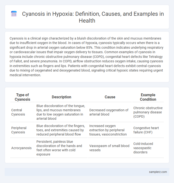
| Type of Cyanosis | Description | Cause | Example Condition |
|---|---|---|---|
| Central Cyanosis | Blue discoloration of the tongue, lips, and mucous membranes due to low oxygen saturation in arterial blood | Decreased oxygenation of arterial blood | Chronic obstructive pulmonary disease (COPD) |
| Peripheral Cyanosis | Blue discoloration of the fingers, toes, and extremities caused by reduced peripheral blood flow | Increased oxygen extraction by peripheral tissues, vasoconstriction | Congestive heart failure (CHF) |
| Acrocyanosis | Persistent, painless blue discoloration of the hands and feet often worse with cold exposure | Vasospasm of small blood vessels | Cold-induced vasospastic disorders |
Understanding Cyanosis: A Key Indicator of Hypoxia
Cyanosis, characterized by a bluish discoloration of the skin and mucous membranes, serves as a critical clinical indicator of hypoxia, reflecting reduced oxygen saturation in the blood. Peripheral cyanosis occurs due to vasoconstriction and reduced blood flow in extremities, while central cyanosis indicates more severe systemic hypoxemia affecting vital organs. Recognizing cyanosis promptly aids healthcare providers in diagnosing underlying respiratory or circulatory conditions that impair oxygen delivery to tissues.
What Is Hypoxia and How Does Cyanosis Manifest?
Hypoxia is a medical condition characterized by insufficient oxygen supply to body tissues, leading to impaired cellular function. Cyanosis manifests as a bluish discoloration of the skin and mucous membranes, particularly visible in the lips, fingertips, and nail beds, indicating decreased oxygen saturation in the blood. This physical sign often signals severe hypoxia requiring immediate clinical assessment and intervention.
Central Cyanosis: Signs and Common Causes in Hypoxia
Central cyanosis in hypoxia presents as a bluish discoloration of the lips, tongue, and mucous membranes, indicating inadequate oxygenation of arterial blood. Common causes include chronic obstructive pulmonary disease (COPD), congenital heart defects with right-to-left shunting, and severe pneumonia, all impairing oxygen delivery to tissues. Early recognition of central cyanosis is crucial for prompt intervention to prevent tissue hypoxia and organ damage.
Peripheral Cyanosis: When Extremities Turn Blue
Peripheral cyanosis occurs in hypoxia when oxygen saturation drops below 85%, causing extremities such as fingers, toes, and lips to develop a bluish discoloration due to reduced peripheral blood oxygen levels. This condition often indicates poor circulation or respiratory issues like chronic obstructive pulmonary disease (COPD) or heart failure, where oxygen delivery to peripheral tissues is compromised. Clinical detection of peripheral cyanosis aids in diagnosing underlying hypoxic states, emphasizing the importance of pulse oximetry and arterial blood gas analysis for accurate assessment.
Cyanosis in Chronic Obstructive Pulmonary Disease (COPD)
Cyanosis in Chronic Obstructive Pulmonary Disease (COPD) manifests as a bluish discoloration of the skin and mucous membranes due to insufficient oxygen saturation in the blood. This hypoxic condition occurs because COPD impairs airflow, leading to chronic low oxygen levels and increased deoxyhemoglobin concentration. Observation of central cyanosis, particularly on the lips and tongue, serves as a crucial clinical indicator of advanced hypoxia in COPD patients.
Congenital Heart Disease and Cyanotic Episodes
Cyanosis in hypoxia commonly manifests in patients with congenital heart disease, particularly those experiencing cyanotic episodes due to right-to-left shunting of deoxygenated blood. Conditions like Tetralogy of Fallot lead to reduced oxygen saturation and characteristic bluish discoloration of the skin and mucous membranes. Monitoring oxygen levels and timely surgical intervention are critical to managing cyanotic episodes and preventing severe complications.
Recognizing Cyanosis in Acute Respiratory Distress
Cyanosis in acute respiratory distress typically presents as a bluish discoloration of the lips, tongue, and nail beds due to inadequate oxygenation of hemoglobin in the blood. Recognizing peripheral cyanosis requires careful assessment under natural light and may signal critical hypoxemia requiring immediate intervention. Clinical signs of central cyanosis, including dusky mucous membranes, are key indicators for prompt airway management and supplemental oxygen therapy.
Cyanosis from High Altitude Hypoxia: What to Look For
Cyanosis from high altitude hypoxia typically presents as a bluish discoloration of the lips, fingers, and toes due to decreased oxygen saturation in the blood caused by lower atmospheric oxygen levels. Symptoms often include shortness of breath, rapid heartbeat, and fatigue, which worsen with exertion at elevations above 2,500 meters. Recognizing this condition early is crucial for preventing severe complications such as high-altitude pulmonary edema or cerebral edema.
When Hypothermia Leads to Cyanotic Symptoms
Hypothermia causes blood vessels to constrict, reducing oxygen delivery to peripheral tissues and leading to cyanosis, particularly in the fingers, toes, and lips. This condition results from decreased oxygen saturation in the hemoglobin, manifesting as a bluish discoloration of the skin. Hypoxia-induced cyanosis in hypothermic patients serves as a critical diagnostic indicator requiring prompt medical intervention to restore adequate tissue oxygenation.
Clinical Assessment: Differentiating Cyanosis Types in Hypoxic Patients
Clinical assessment of cyanosis in hypoxic patients involves distinguishing between central and peripheral cyanosis based on skin and mucous membrane coloration. Central cyanosis presents as a bluish discoloration of the lips and tongue due to decreased arterial oxygen saturation, while peripheral cyanosis typically affects the extremities and is caused by slowed blood flow or vasoconstriction. Pulse oximetry and arterial blood gas analysis are essential diagnostic tools for accurately evaluating oxygen saturation levels and guiding appropriate treatment strategies.
example of cyanosis in hypoxia Infographic