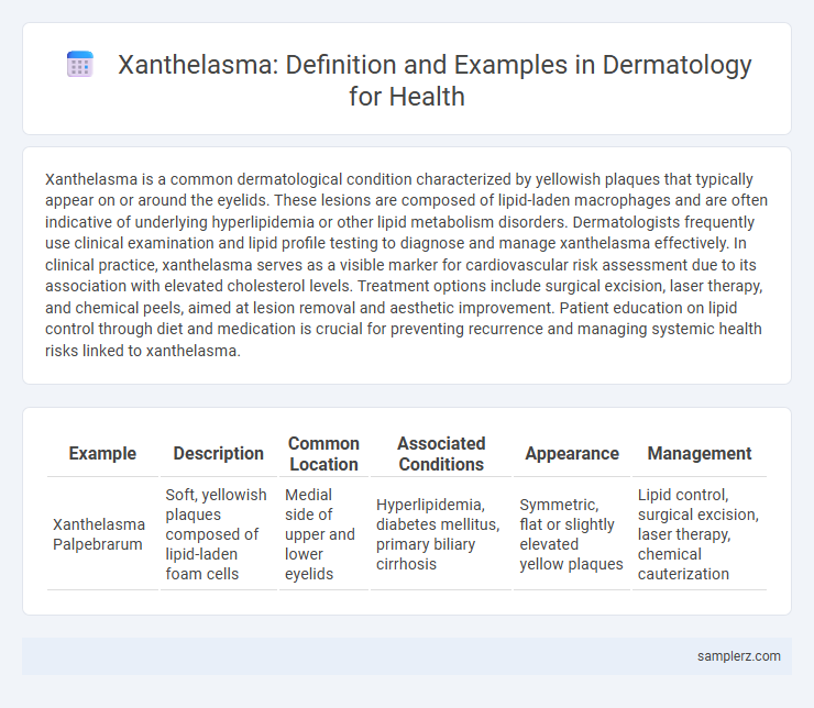
Xanthelasma is a common dermatological condition characterized by yellowish plaques that typically appear on or around the eyelids. These lesions are composed of lipid-laden macrophages and are often indicative of underlying hyperlipidemia or other lipid metabolism disorders. Dermatologists frequently use clinical examination and lipid profile testing to diagnose and manage xanthelasma effectively. In clinical practice, xanthelasma serves as a visible marker for cardiovascular risk assessment due to its association with elevated cholesterol levels. Treatment options include surgical excision, laser therapy, and chemical peels, aimed at lesion removal and aesthetic improvement. Patient education on lipid control through diet and medication is crucial for preventing recurrence and managing systemic health risks linked to xanthelasma.
Table of Comparison
| Example | Description | Common Location | Associated Conditions | Appearance | Management |
|---|---|---|---|---|---|
| Xanthelasma Palpebrarum | Soft, yellowish plaques composed of lipid-laden foam cells | Medial side of upper and lower eyelids | Hyperlipidemia, diabetes mellitus, primary biliary cirrhosis | Symmetric, flat or slightly elevated yellow plaques | Lipid control, surgical excision, laser therapy, chemical cauterization |
Understanding Xanthelasma: Definition and Clinical Presentation
Xanthelasma is a dermatological condition characterized by yellowish, soft plaques typically located on the eyelids, especially near the inner canthus. These lipid-rich deposits are composed primarily of cholesterol-laden foam cells within the dermis and often indicate underlying hyperlipidemia. Clinically, xanthelasma presents as asymptomatic, flat or slightly elevated lesions that may gradually increase in size and number, warranting evaluation for cardiovascular risk factors.
Epidemiology and Prevalence of Xanthelasma
Xanthelasma, characterized by yellowish plaques on the eyelids, primarily affects individuals aged 40 to 60 years, with a higher prevalence in females. Epidemiological studies reveal a global prevalence ranging from 0.56% to 1.5%, often associated with dyslipidemia and metabolic syndrome. The condition is more commonly observed in populations with hyperlipidemia, emphasizing the importance of lipid profile screening in dermatological assessments.
Common Sites and Morphology of Xanthelasma Lesions
Xanthelasma lesions typically appear as soft, yellowish plaques located on the medial aspects of the eyelids, especially near the inner canthus. These lesions are flat or slightly elevated with well-defined edges and can vary in size from a few millimeters to several centimeters. Common sites also include the upper eyelids more frequently than the lower eyelids, with symmetry often observed bilaterally.
Xanthelasma vs. Other Cutaneous Xanthomas
Xanthelasma presents as yellowish plaques predominantly around the eyelids and is the most common form of cutaneous xanthomas, often associated with lipid metabolism disorders. Unlike eruptive xanthomas, which appear as multiple small, yellow papules on the trunk and extremities linked to hypertriglyceridemia, xanthelasma is more localized and less commonly related to severe lipid abnormalities. Identifying and differentiating xanthelasma from other xanthomas is crucial for assessing underlying systemic conditions such as hyperlipidemia or diabetes mellitus.
Risk Factors and Associated Systemic Conditions
Xanthelasma presents as yellowish plaques typically located on the eyelids, often linked to lipid metabolism disorders such as hyperlipidemia and familial hypercholesterolemia. Risk factors include advancing age, diabetes mellitus, and hypothyroidism, which contribute to altered lipid profiles and increase lesion development. This dermatologic manifestation is frequently associated with systemic conditions like cardiovascular disease and metabolic syndrome, underscoring the importance of comprehensive lipid screening in affected patients.
Histopathological Features of Xanthelasma
Xanthelasma histopathology reveals foam cells laden with lipid material within the dermis, primarily in the perivascular and periadnexal areas. The lesion consists of clusters of multivacuolated histiocytes embedded among collagen fibers, often accompanied by mild inflammation. Cholesterol clefts and extracellular lipid deposits are also characteristic microscopic findings in xanthelasma specimens.
Diagnostic Approaches in Identifying Xanthelasma
Xanthelasma is identified primarily through clinical examination, where characteristic yellowish plaques typically appear on the eyelids. Dermoscopy enhances diagnostic accuracy by revealing lipid-rich deposits with a distinctive pattern, aiding differentiation from other eyelid lesions. Biopsy and histopathological analysis confirm diagnosis by demonstrating foam cells within the dermis, essential for cases with atypical presentation or suspected malignancy.
Case Examples of Xanthelasma in Dermatological Practice
Xanthelasma appears as yellowish plaques commonly located on the medial eyelids, often indicating lipid metabolism disorders. Case studies reveal patients with xanthelasma frequently exhibit elevated serum cholesterol and triglyceride levels, underscoring the need for lipid profile evaluations. Dermatological practice emphasizes differential diagnosis to distinguish xanthelasma from conditions like syringoma or sebaceous hyperplasia, ensuring accurate treatment planning.
Management and Treatment Options for Xanthelasma
Xanthelasma, characterized by yellowish cholesterol-rich plaques around the eyelids, requires careful management to prevent recurrence and minimize cosmetic concerns. Treatment options include surgical excision, laser therapy such as CO2 or erbium lasers, and chemical peels with trichloroacetic acid, each offering varying efficacy based on lesion size and patient skin type. Lifestyle modifications and lipid-lowering medications like statins are recommended to address underlying hyperlipidemia and reduce the risk of new xanthelasma formation.
Prevention and Prognosis of Xanthelasma in Patients
Xanthelasma, characterized by yellowish plaques on the eyelids, often signals underlying lipid metabolism disorders, making lipid profile management crucial for prevention. Maintaining healthy cholesterol and triglyceride levels through diet, exercise, and medication reduces recurrence risk and progression of xanthelasma. Prognosis remains favorable with early intervention, although lesions may persist cosmetically without treatment.

example of xanthelasma in dermatology Infographic
 samplerz.com