Sarcoidosis is a systemic inflammatory disease characterized by the formation of non-caseating granulomas, which are clusters of immune cells. These granulomas primarily affect the lungs and lymph nodes, but can also involve organs like the skin, eyes, and liver. The granulomas consist of tightly packed macrophages, epithelioid cells, and multinucleated giant cells, indicating an immune response to an unknown antigen. In sarcoidosis, granulomas disrupt normal tissue architecture, leading to impaired organ function over time. Diagnosis often relies on biopsy samples revealing the presence of these distinctive granulomas without necrosis. Observation of granuloma formation in sarcoidosis aids in differentiating it from other granulomatous diseases such as tuberculosis, where caseating granulomas are present.
Table of Comparison
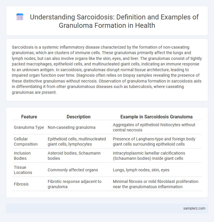
| Feature | Description | Example in Sarcoidosis Granuloma |
|---|---|---|
| Granuloma Type | Non-caseating granuloma | Aggregates of epithelioid histiocytes without central necrosis |
| Cellular Composition | Epithelioid cells, multinucleated giant cells, lymphocytes | Presence of Langhans-type and foreign body giant cells surrounding epithelioid cells |
| Inclusion Bodies | Asteroid bodies, Schaumann bodies | Intracytoplasmic lamellar calcifications (Schaumann bodies) inside giant cells |
| Tissue Locations | Commonly affected organs | Lungs, lymph nodes, skin, eyes |
| Fibrosis | Fibrotic response adjacent to granuloma | Minimal fibrosis or mild fibroblast proliferation near the granulomatous inflammation |
Overview of Sarcoidosis and Granuloma Formation
Sarcoidosis is an inflammatory disease characterized by the formation of non-caseating granulomas, primarily in the lungs, lymph nodes, and skin. Granuloma formation in sarcoidosis involves the aggregation of macrophages, epithelioid cells, and multinucleated giant cells attempting to isolate persistent antigens or unknown triggers. This immune response leads to tissue inflammation and fibrosis, impacting organ function and necessitating diagnostic evaluation through imaging and biopsy.
Pathophysiology of Sarcoid Granulomas
Sarcoid granulomas form due to an exaggerated immune response involving activated T-helper cells and macrophages, leading to non-caseating granulomatous inflammation. Cytokines such as tumor necrosis factor-alpha (TNF-a) and interferon-gamma (IFN-g) drive granuloma formation by promoting macrophage aggregation and differentiation into epithelioid and multinucleated giant cells. These granulomas primarily target the lungs and lymphatic system, disrupting normal tissue architecture and causing clinical manifestations of sarcoidosis.
Key Histological Features of Sarcoid Granulomas
Sarcoid granulomas are characterized by tightly clustered epithelioid histiocytes with minimal central necrosis, commonly exhibiting multinucleated giant cells such as Langhans-type and asteroid bodies. These non-caseating granulomas prominently lack the extensive lipid necrosis seen in tuberculosis, with sparse surrounding lymphocytic infiltration forming a "naked" granuloma appearance. The presence of fibroblasts at the periphery indicates early fibrosis, a hallmark of chronic sarcoidosis tissue response.
Typical Organs Affected by Sarcoid Granulomas
Sarcoid granulomas typically affect the lungs, lymph nodes, skin, and eyes, with pulmonary involvement seen in over 90% of cases. These granulomas consist of tightly clustered immune cells that cause inflammation and tissue damage, leading to symptoms like chronic cough, shortness of breath, and skin lesions. Other organs such as the liver, heart, and nervous system may also be involved but less frequently.
Pulmonary Sarcoidosis: Lung Granuloma Examples
Pulmonary sarcoidosis primarily manifests through the formation of non-caseating granulomas in lung tissue, which disrupt normal respiratory function. These granulomas consist of tightly clustered immune cells, including epithelioid histiocytes and multinucleated giant cells, surrounded by lymphocytes. High-resolution CT scans often reveal bilateral hilar lymphadenopathy and fine nodular infiltrates characteristic of lung granulomas in sarcoidosis patients.
Cutaneous Manifestations: Skin Granulomas in Sarcoidosis
Cutaneous manifestations of sarcoidosis often present as non-caseating granulomas within the skin, characterized by firm, reddish-brown papules, plaques, or nodules commonly found on the face, neck, and extremities. Histopathological examination reveals tightly clustered epithelioid cells and multinucleated giant cells without central necrosis, distinguishing sarcoid granulomas from infectious granulomas. These skin granulomas reflect systemic involvement and can aid in diagnosing sarcoidosis alongside pulmonary and lymph node findings.
Ocular Sarcoidosis: Eye Granuloma Presentation
Ocular sarcoidosis commonly presents with granulomatous uveitis, characterized by the formation of non-caseating granulomas in the eye's uveal tract. These granulomas often lead to symptoms such as blurred vision, photophobia, and redness, with typical involvement of the iris and conjunctiva. Diagnosis relies on clinical examination, supported by imaging techniques like slit-lamp biomicroscopy and anterior segment optical coherence tomography (OCT), to visualize characteristic granulomatous inflammation.
Cardiac Involvement: Heart Granulomas in Sarcoidosis
Sarcoidosis frequently causes granulomas in cardiac tissue, leading to conduction abnormalities, arrhythmias, and heart failure. The presence of non-caseating granulomas disrupts normal myocardial function and can cause sudden cardiac death in severe cases. Cardiac magnetic resonance imaging (MRI) and endomyocardial biopsy are essential for diagnosing heart granulomas in sarcoidosis patients.
Diagnostic Imaging of Sarcoid Granulomas
Sarcoid granulomas commonly appear on chest X-rays as bilateral hilar lymphadenopathy and pulmonary nodules, which are key diagnostic imaging features. High-resolution computed tomography (HRCT) provides detailed visualization of granulomatous inflammation, revealing perilymphatic nodules and fibrosis patterns characteristic of sarcoidosis. Positron emission tomography (PET) scans can detect metabolically active granulomas, aiding in disease staging and monitoring therapeutic response.
Differentiating Sarcoid Granulomas from Other Granulomatous Diseases
Sarcoid granulomas characteristically present as non-caseating, compact clusters of epithelioid histiocytes with multinucleated giant cells, distinguishing them from infectious granulomas that typically exhibit caseous necrosis. The absence of central necrosis and the presence of asteroid or Schaumann bodies within giant cells serve as key histopathological markers favoring sarcoidosis. Differential diagnosis requires correlating clinical, radiological, and laboratory findings, including serum angiotensin-converting enzyme levels and exclusion of tuberculosis or fungal infections.
example of sarcoidosis in granuloma Infographic