Erythema nodosum is a skin condition characterized by tender, red nodules usually appearing on the shins. These nodules often measure between 1 to 5 centimeters in diameter and can be accompanied by swelling and warmth in the affected area. The condition is commonly associated with infections, medications, or autoimmune diseases, which trigger inflammation in the fat cells under the skin. In a typical case affecting the leg, erythema nodosum presents as symmetrical, raised lesions that may be painful to touch. Patients might experience systemic symptoms such as fever, joint pain, and fatigue along with these skin manifestations. Diagnosing erythema nodosum involves clinical examination and sometimes biopsy to confirm the presence of septal panniculitis without vasculitis.
Table of Comparison
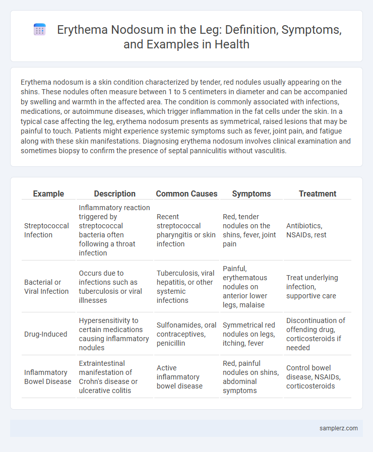
| Example | Description | Common Causes | Symptoms | Treatment |
|---|---|---|---|---|
| Streptococcal Infection | Inflammatory reaction triggered by streptococcal bacteria often following a throat infection | Recent streptococcal pharyngitis or skin infection | Red, tender nodules on the shins, fever, joint pain | Antibiotics, NSAIDs, rest |
| Bacterial or Viral Infection | Occurs due to infections such as tuberculosis or viral illnesses | Tuberculosis, viral hepatitis, or other systemic infections | Painful, erythematous nodules on anterior lower legs, malaise | Treat underlying infection, supportive care |
| Drug-Induced | Hypersensitivity to certain medications causing inflammatory nodules | Sulfonamides, oral contraceptives, penicillin | Symmetrical red nodules on legs, itching, fever | Discontinuation of offending drug, corticosteroids if needed |
| Inflammatory Bowel Disease | Extraintestinal manifestation of Crohn's disease or ulcerative colitis | Active inflammatory bowel disease | Red, painful nodules on shins, abdominal symptoms | Control bowel disease, NSAIDs, corticosteroids |
Overview of Erythema Nodosum in the Leg
Erythema nodosum in the leg presents as tender, red nodules typically located on the anterior aspects of the lower legs. This inflammatory condition involves the subcutaneous fat and is often associated with infections, medications, or systemic diseases like sarcoidosis. Diagnosis relies on clinical examination supported by biopsy when needed, and treatment targets the underlying cause along with symptomatic relief.
Common Causes of Erythema Nodosum in Lower Limbs
Erythema nodosum in the lower limbs most commonly results from infections such as streptococcal pharyngitis, tuberculosis, and fungal diseases like coccidioidomycosis. Other frequent causes include inflammatory conditions like sarcoidosis and inflammatory bowel diseases, including Crohn's disease and ulcerative colitis. Drug reactions, particularly to antibiotics and oral contraceptives, also represent significant triggers for erythema nodosum in the legs.
Recognizing Symptoms of Leg Erythema Nodosum
Painful, tender red nodules typically appear on the front of the lower legs, often accompanied by swelling and warmth in the affected area. Patients may also experience fever, fatigue, and joint pain, which signal an underlying inflammatory response. Early recognition of these distinctive, raised lesions helps in prompt diagnosis and management of leg erythema nodosum.
Visual Characteristics: Erythema Nodosum Lesions on Legs
Erythema nodosum lesions on the legs present as tender, erythematous nodules primarily located on the anterior tibial surfaces. These raised, red to purple bumps often measure 1 to 5 centimeters in diameter and may coalesce into larger plaques. The lesions typically exhibit a symmetrical distribution and can be accompanied by swelling and warmth, indicating underlying inflammation.
Differential Diagnosis for Leg Nodules
Erythema nodosum commonly presents as tender, red nodules on the anterior aspects of the legs, often linked to infections, medications, or systemic diseases like sarcoidosis and inflammatory bowel disease. Differential diagnosis for leg nodules includes cellulitis, vasculitis, lipomas, and deep vein thrombosis, each distinguished by clinical evaluation and diagnostic imaging. Accurate diagnosis relies on histopathological examination and correlating clinical features to exclude other causes such as panniculitis or malignancies.
Systemic Conditions Associated with Erythema Nodosum
Erythema nodosum commonly presents as tender, red nodules on the shins and is often associated with systemic conditions such as streptococcal infections, sarcoidosis, inflammatory bowel disease, and tuberculosis. These underlying diseases trigger a hypersensitivity reaction leading to inflammation of the subcutaneous fat. Recognizing erythema nodosum as a clinical sign can prompt early investigation and management of its systemic causes.
Diagnostic Approach for Leg Erythematous Nodules
Diagnostic approach for leg erythematous nodules includes a thorough clinical examination focusing on the characteristic tender, red, and warm nodules typically located on the anterior shins. Laboratory tests such as complete blood count, erythrocyte sedimentation rate (ESR), and C-reactive protein (CRP) help assess inflammation, while skin biopsy confirms erythema nodosum by revealing septal panniculitis without vasculitis. Additional investigations like chest X-ray or throat culture may be necessary to identify underlying causes such as infections, sarcoidosis, or autoimmune diseases.
Treatment Options for Erythema Nodosum in the Leg
Treatment options for erythema nodosum in the leg primarily include nonsteroidal anti-inflammatory drugs (NSAIDs) such as ibuprofen to reduce pain and inflammation. In severe or persistent cases, corticosteroids like prednisone may be prescribed to control the immune response. Supportive measures such as leg elevation, compression stockings, and rest are also effective in alleviating symptoms and promoting healing.
Prognosis and Recovery: What to Expect
Erythema nodosum on the leg typically resolves within 3 to 6 weeks with appropriate treatment, and most patients experience full recovery without scarring. The prognosis is generally excellent, though recurrent episodes can occur, especially if the underlying cause remains untreated. Recovery includes gradual fading of painful red nodules and restoration of normal skin texture and function.
Patient Case Example: Erythema Nodosum Affecting the Leg
A 28-year-old female patient presented with tender, erythematous nodules on the anterior aspect of her lower legs, consistent with erythema nodosum. Laboratory tests revealed elevated inflammatory markers and a recent streptococcal infection, common triggers of this panniculitis. Treatment included nonsteroidal anti-inflammatory drugs and addressing the underlying infection, resulting in gradual lesion resolution over four weeks.
example of erythema nodosum in leg Infographic