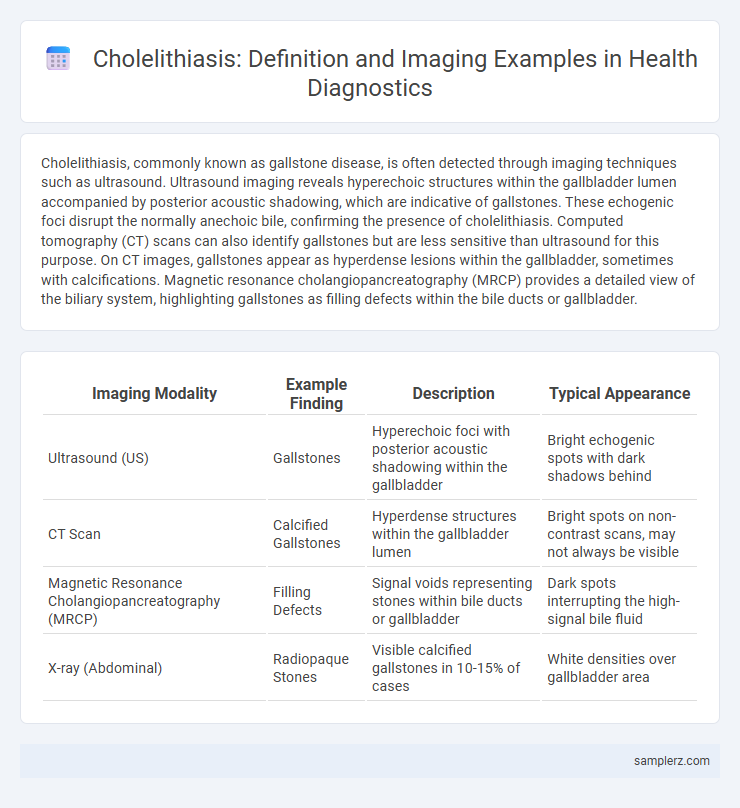
Cholelithiasis, commonly known as gallstone disease, is often detected through imaging techniques such as ultrasound. Ultrasound imaging reveals hyperechoic structures within the gallbladder lumen accompanied by posterior acoustic shadowing, which are indicative of gallstones. These echogenic foci disrupt the normally anechoic bile, confirming the presence of cholelithiasis. Computed tomography (CT) scans can also identify gallstones but are less sensitive than ultrasound for this purpose. On CT images, gallstones appear as hyperdense lesions within the gallbladder, sometimes with calcifications. Magnetic resonance cholangiopancreatography (MRCP) provides a detailed view of the biliary system, highlighting gallstones as filling defects within the bile ducts or gallbladder.
Table of Comparison
| Imaging Modality | Example Finding | Description | Typical Appearance |
|---|---|---|---|
| Ultrasound (US) | Gallstones | Hyperechoic foci with posterior acoustic shadowing within the gallbladder | Bright echogenic spots with dark shadows behind |
| CT Scan | Calcified Gallstones | Hyperdense structures within the gallbladder lumen | Bright spots on non-contrast scans, may not always be visible |
| Magnetic Resonance Cholangiopancreatography (MRCP) | Filling Defects | Signal voids representing stones within bile ducts or gallbladder | Dark spots interrupting the high-signal bile fluid |
| X-ray (Abdominal) | Radiopaque Stones | Visible calcified gallstones in 10-15% of cases | White densities over gallbladder area |
Overview of Cholelithiasis and Its Clinical Significance
Cholelithiasis, characterized by the presence of gallstones within the gallbladder, is commonly identified through imaging modalities such as abdominal ultrasound, which reveals echogenic foci with posterior acoustic shadowing. The clinical significance of cholelithiasis lies in its potential to cause biliary colic, cholecystitis, and complications like choledocholithiasis or pancreatitis, requiring prompt diagnosis and management. Imaging findings guide treatment decisions, including conservative monitoring or surgical intervention like cholecystectomy.
Common Imaging Modalities for Cholelithiasis Detection
Ultrasound remains the primary imaging modality for detecting cholelithiasis due to its high sensitivity in visualizing gallstones as echogenic foci with posterior acoustic shadowing. Computed tomography (CT) can identify calcified gallstones and assess complications such as cholecystitis, although it is less sensitive for non-calcified stones. Magnetic resonance cholangiopancreatography (MRCP) provides detailed visualization of the biliary tree and is effective for detecting gallstones within the common bile duct.
Ultrasonography Findings in Cholelithiasis
Ultrasonography in cholelithiasis typically reveals echogenic foci with posterior acoustic shadowing within the gallbladder lumen, indicative of gallstones. The gallbladder wall may appear thickened, often exceeding 3mm, suggesting inflammation or cholecystitis. Sonographic Murphy's sign, characterized by focal tenderness over the gallbladder during ultrasound probe pressure, supports the diagnosis of symptomatic gallstones.
CT Scan Features of Gallstones
Gallstones appear as hyperdense foci within the gallbladder lumen on CT scans, often causing acoustic shadowing. Radiologists identify well-defined, high-attenuation lesions that may vary in size and number, with some gallstones exhibiting calcifications visible on non-contrast CT. CT imaging enhances detection of complications like gallbladder wall thickening or pericholecystic fluid, critical for diagnosing acute cholecystitis associated with cholelithiasis.
MRI and MRCP Appearance in Cholelithiasis Cases
Magnetic Resonance Imaging (MRI) and Magnetic Resonance Cholangiopancreatography (MRCP) demonstrate cholelithiasis with characteristic findings such as signal voids within the gallbladder lumen representing gallstones, often appearing as low-intensity foci on T2-weighted images against a high-signal bile background. MRCP provides detailed visualization of the biliary tree, revealing filling defects corresponding to stones within the cystic duct or common bile duct, aiding in the diagnosis of choledocholithiasis. These imaging modalities are critical for non-invasive assessment of gallstone burden, bile duct obstruction, and associated complications like cholecystitis.
Endoscopic Retrograde Cholangiopancreatography (ERCP) in Gallstone Evaluation
Endoscopic Retrograde Cholangiopancreatography (ERCP) provides detailed imaging of the biliary tree, making it essential for diagnosing cholelithiasis, particularly in detecting gallstones within the common bile duct. This technique combines endoscopy and fluoroscopy to visualize and allows for therapeutic interventions such as stone extraction or stent placement. ERCP's high sensitivity and specificity enhance the evaluation of choledocholithiasis, guiding effective clinical management.
Typical Imaging Signs: Sludge, Shadowing, and Mobile Stones
Cholelithiasis typically appears on ultrasound imaging as echogenic foci with posterior acoustic shadowing, indicative of gallstones. Gallbladder sludge presents as low-level, non-shadowing echoes that layer dependently without acoustic shadowing. Mobile stones shift position with patient movement, helping differentiate them from polyps or tumors in the gallbladder lumen.
Differentiating Cholelithiasis from Other Biliary Pathologies
Cholelithiasis is characterized by echogenic foci with posterior acoustic shadowing on ultrasound, distinct from biliary sludge which lacks shadowing and appears as low-level echoes without mobility. On CT imaging, gallstones appear as hyperdense structures within the gallbladder lumen, whereas biliary duct strictures present as localized narrowing without intraluminal calcifications. MRI with MRCP highlights filling defects within the bile ducts for choledocholithiasis, contrasting with wall thickening or enhancement seen in cholangitis and biliary tumors.
Case Studies: Classic Imaging Examples of Gallstones
Cholelithiasis commonly presents in imaging studies such as abdominal ultrasound, where hyperechoic foci with posterior acoustic shadowing in the gallbladder lumen indicate gallstones. Case studies often highlight the use of CT scans revealing hyperdense calculi within the gallbladder, sometimes accompanied by gallbladder wall thickening indicative of inflammation. Magnetic resonance cholangiopancreatography (MRCP) provides detailed visualization of biliary obstruction caused by gallstones, essential for diagnosis and treatment planning.
Importance of Early Imaging Diagnosis in Cholelithiasis Management
Early imaging diagnosis of cholelithiasis using ultrasound reveals hyperechoic gallstones with acoustic shadowing, facilitating timely intervention and preventing complications such as cholecystitis or pancreatitis. Computed tomography (CT) and magnetic resonance cholangiopancreatography (MRCP) provide detailed visualization of biliary obstruction and stone location, guiding surgical planning. Prompt detection through imaging significantly reduces morbidity and enhances patient outcomes in cholelithiasis management.
example of cholelithiasis in imaging Infographic